Just another iHealthSpot WP02 site

Orion Physical Therapy

  • Lake Orion(248) 814-8060
  • Midland(989) 832-9300
Active Orthopedics - Physical Therapy
MENUMENU
  • Home
  • Meet Our Teams
    • Lake Orion
    • Midland
  • Services
    • Manual Therapy
    • Pre-and Post-Surgical Rehabilitation
    • Balance Disorders and Gait Training
    • Sports Injuries
    • Work Injuries
  • Special Programs
    • Lake Orion Programs
    • Midland Programs
  • Patient Forms
  • Locations
  • Contact Us

Pediatrics - Fractures

Back to Patient Education
  • Introduction
  • Anatomy
  • Causes
  • Symptoms
  • Diagnosis
  • Surgery
  • Treatment
  • Recovery
  • Prevention

Introduction

Bones are the hardest tissues in the body.  Although bones are strong, they can split or break under too much pressure or force.  A broken bone is called a fracture.  Fractures can occur in a variety of ways.  The most common causes of fractures are injuries, prolonged stress from overuse, and bone weakening diseases, such as Osteoporosis or tumors. 

There are many types of fractures.  They can range from a hairline crack to a bone that has broken into several pieces.  Simple fractures may only require casting or splinting treatments.  More complex fractures may need surgical intervention to align the bones for proper healing.

Back to top

Anatomy

Your child’s bones vary in size and shape.  For instance, your child’s arms and legs contain long bones.  A series of small bones, called vertebrae, make up your child’s spine.  Very small bones form your child’s hands and feet.  Some of your child’s bones have curves, including his or her ribs and skull.  All of your child’s bones line up and connect to form his or her skeleton.  In addition to creating your child’s body structure, your child’s bones produce blood cells, form joints with muscles for movement, and protect internal organs. 
 
Your child’s bones are live tissues.  They change and grow like the other parts of your child’s body.  Most of the bones in your child’s body are composed of the same layered materials. 
 
The outer layer of a bone is called the periosteum.  It is considered the life support system for the bone because it provides the nutrient blood for bone cells.  The periosteum also produces bone-developing cells during growth or after a fracture.  Underneath the periosteum is compact bone, known as the cortex.  Compact bone is solid and hard.  It covers the cancellous “spongy” bone.  The cancellous bone looks like a sponge because it contains many pores.  It can resist the stresses of weight, postural changes, and growth.  In many bones, the cancellous bone contains or protects the red marrow or bone marrow.  Red marrow contains developing and mature blood cells.
Back to top

Causes

The most frequent causes of fractures are falls and motor vehicle crashes.  There is a higher incidence of fractures in some sports that involve prolonged impact, high impact, balance, or high speeds.  Stress fractures can result from prolonged impact or repetitive forces.  For example, running or jogging can cause stress fractures in the leg, foot, ankle, or hip.  High impacts can occur during contact sports, including tackles in football or punches in boxing.  Skateboarding, bicycle riding, and snow skiing are sports that involve balance and speed.  Fractures can occur during contact with a hard surface, for instance during a fall to the cement while skateboarding or during contact with an object, such as a tree while snow skiing.
 
Fractures can also be the result of physical violence.  Fractures can result from a blow with a fist or kick, or from contact with a solid weapon, such as a bat.
 
Although the majority of fractures result from motor vehicle crashes and falls, some fractures occur because of diseases.  Osteoporosis is a medical condition that causes more bone calcium to be absorbed than is replaced.  Calcium is necessary for hard healthy bones.  Osteoporosis causes a reduction in bone density and brittle or fragile bones that are vulnerable to fractures.  Type I Osteoporosis usually affects women between the ages of 51 and 75.  Type I Osteoporosis is associated with spine and wrist fractures.  Type II Osteoporosis usually affects people between the ages of 70 and 85.  Type II Osteoporosis is associated with hip, pelvis, arm, and leg fractures.
 
Bone tumors are another disease-related cause of fractures.  Most bone tumors originate elsewhere in the body and metastasize or spread to the bone.  Very rarely do cancerous tumors begin in the bone.  Tumors can weaken bones, making them susceptible to fractures.
Back to top

Symptoms

In some cases, a snap or cracking sound may be heard when a bone fractures.  Your child may feel sharp, deep, or intense pain along with numbness or tingling.  Your child’s skin may swell, bruise, or bleed.
 
The place where your child’s fracture occurs may look odd, bent, or out of place.  Sometimes a broken bone may come through the skin.  Your child may not be able to move or put weight on his or her limb or joint, or he or she may do so with difficulty.
Back to top

Diagnosis

Your doctor can diagnose a fracture with a physical examination.  Your doctor will ask you to describe your child’s injury and your symptoms.  In most cases, imaging tests are ordered to confirm the fracture.
 
An X-ray will be ordered to identify the type and location of your child’s fracture.  Some fractures, such as stress fractures, may not show up on an X-ray.  In such cases, Computed Tomography (CT) scans or Magnetic Resonance Imaging (MRI) scans may be used to take a more detailed look at your child’s bones.  X-rays, CT scans, and MRI scans are painless procedures.
 
A bone scan is useful for identifying bone abnormalities from Osteoporosis or cancer.  A bone scan may be used to show fractures, tumors, infection, and bone deterioration.  A bone scan requires that your child receive a small harmless injection of a radioactive substance several hours before your test.  The substance collects in areas where the bone is breaking down or repairing itself.
 
In addition to diagnosing your fracture, your doctor will classify the type of fracture that your child has in order to plan treatment appropriately.  Fractures are classified by a combination of general terms used to describe their features.  Fractures are categorized by the characteristics of the broken bone, including the position of the fragments or broken bone and the direction of the fracture line.  Common fracture characteristics and classifications are described below.
 
A fracture is first classified in general terms:
 
Complete Fracture: The bone is completely broken into separate pieces.
Incomplete or Partial Fracture: A crack that does not completely break the bone into two pieces.
Greenstick Fracture:  An Incomplete Fracture with a bowed bone, it is more common in children.
Compound or Open Fracture: The bone fragments penetrate the skin.
Simple or Closed Fracture: The bone fragments do not penetrate the skin.
 
Fractures are further described and classified by the position of the bone fragments:
 
Comminuted: The bones are broken into several pieces.
Nondisplaced: The bone is broken but maintains its alignment.
Displaced: The bone is broken and the fragments are out of position.
Segmental:  More than one fracture line leads to a "floating" segment.
Angulated: The fragments are out of position and at an angle to each other.
Overriding: The fragments overlap each other.
Impacted: One piece of bone is forced into a second piece of bone.
 
The fracture line or crack is also described and classified.  This terminology is especially used to describe fractures in the long bones of the arms and legs:
 
Linear: The fracture line is parallel with the shaft (the long part) of the bone.
Transverse:  The fracture line is at a right (90°) angle to the shaft of the bone.
Oblique:  The fracture line is at a 45° angle to the shaft of the bone.
Spiral:  The fracture line has a “corkscrewed,” “curled” or angled pattern.
Back to top

Surgery

Surgery is recommended for fractures that do not heal properly or when the bones have broken in such a way that they are unlikely to remain aligned when set with a cast.  There are several options for surgery.  The type of surgery that your child receives will depend on the location and type of your child’s fracture.  Your child can have general anesthesia for surgery or your doctor can numb the area with a nerve block.
 
Surgical options include procedures called an Open Reduction and Internal Fixation or an Open Reduction and External Fixation.  Open Reduction and Internal Fixation refers to techniques that use surgical hardware to stabilize a fracture beneath the skin.  Your surgeon will make an incision and place your child’s bones in the proper position for healing.  Your surgeon will secure the bones together with surgical hardware, such as rods, screws, or metal plates. 
 
Open Reduction and External Fixation refers to techniques that use surgical hardware to stabilize a fracture from the outside of the skin.  Your surgeon will make an incision and place your child’s bones in the proper position for healing.  Your surgeon will secure the bones with surgical pins that are placed through the outside of the skin.  The surgical pins are attached to a metal frame on the outside of the skin. 
Back to top

Treatment

The treatment for a fracture depends on the type of fracture and the bones that are involved.  The goal of treatment is to align the fractured bones to allow them to heal.  A fractured bone is able to grow back together.  When a bone is fractured, it produces a protective blood clot and callus at the end of the bones.  The ends of the bones created new bone cells and blood vessels that grow towards each other and eventually heal the bone.
 
Your doctor will use X-rays as a guide to “set” or position your child’s bones.  Some bones can be kept in place with a cast while they heal.  The cast keeps the bones from moving.  The hard surface of the cast protects the injury.  Casts are made of a variety of materials.  They are usually worn for about one to two months, depending on the type of fracture and the bone involved. 
 
A fracture which does not heal or "unite" within the usual period of time for that fracture is said to have delayed union.  The reason for the delay is not always known, but usually results from excess motion at the fracture during healing or a poor blood supply.  When healing does occur, it has taken longer than normal.
 
A nonunion is a fracture that just will not heal.  This is usually determined by time and X-rays, which show that there is no possibility that the fracture will heal without additional intervention.
 
A malunion is a fracture that has healed in a position outside acceptable parameters.  This can include combination of angulation, displacement, malrotation, or length differences.
Delayed unions, nonunions and malunions generally require additional treatment.  Bone stimulators may be used to enhance fracture healing.  These are usually used externally, but sometimes are placed surgically at the fracture site.  They use either electricity or ultrasound, which has been shown to enhance bone generation.
 
Malunions and many nonunions require surgery to heal properly.  Direct access to the fracture is necessary to straighten the bone, in the case of a malunion, or to remove soft tissues and stimulate blood flow at the site of a nonunion.  Bone grafts are typically placed to further enhance healing.
 
Your child's pain will probably cease before his or her fracture has completely healed.  Your doctor will limit your child's activity while his or her bone is healing.  An immobilized joint may become stiff and be difficult to move.  Because of this, therapy may follow surgery or casting.  Therapists will work with your child to regain movement, strength, and flexibility that may have decreased while his or her bone or joint was immobile.  The therapists may apply modalities, such as heat or ultrasound, to sooth your child's pain.  Your child's therapists can also recommend safety equipment that your child can wear or use during sports, at school, or during activities at home to support and protect his or her bones and joints.
Back to top

Recovery

Recovery time from a fracture is different for everyone.  It depends on the type of fracture your child had and the type of treatment your child received.  Your doctor will let you know what to expect.  Generally, fractures need about six weeks to heal.  Some fractures can take several months to heal.  Most people have good outcomes with treatment and are able to return to their regular activities.
Back to top

Prevention

There are several things that your child can do to help prevent fractures.  Your child should wear a seatbelt and use the appropriate seating device while riding in a vehicle.
 
Prevent falls.  A general physical examination can identify medical conditions that are associated with balance disorders or dizziness. 
 
A vision exam can detect vision changes that are associated with falls.  Some vision changes can be corrected with glasses. 
 
If your child plays sports, make sure that your child wears the appropriate safety equipment.  Safety helmets, pads, and body gear designed for your child’s sport or activity can help protect his or her body.
 
Teach your child how to keep his or her bones healthy.  Educate your child about not smoking. Smoking can inhibit the healing process of bones.  Make sure your child’s diet contains healthy amounts of calcium and Vitamin D.  Talk to your doctor about nutrition supplements that may be appropriate for your child.
Back to top

Copyright ©  - iHealthSpot Interactive - www.iHealthSpot.com

This information is intended for educational and informational purposes only. It should not be used in place of an individual consultation or examination or replace the advice of your health care professional and should not be relied upon to determine diagnosis or course of treatment.

The iHealthSpot patient education library was written collaboratively by the iHealthSpot editorial team which includes Senior Medical Authors Dr. Mary Car-Blanchard, OTD/OTR/L and Valerie K. Clark, and the following editorial advisors: Steve Meadows, MD, Ernie F. Soto, DDS, Ronald J. Glatzer, MD, Jonathan Rosenberg, MD, Christopher M. Nolte, MD, David Applebaum, MD, Jonathan M. Tarrash, MD, and Paula Soto, RN/BSN. This content complies with the HONcode standard for trustworthy health information. The library commenced development on September 1, 2005 with the latest update/addition on February 16, 2022. For information on iHealthSpot’s other services including medical website design, visit www.iHealthSpot.com.

Footer

Patient Education

From symptoms and diagnosis to treatment and prevention, learn about common acute and chronic conditions that affect the musculoskeletal system in our patient education center.

LEARN MORE

Special Programs

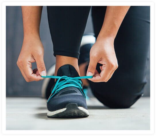
Thanks to advanced certifications our physical therapists have obtained, Orion Physical Therapy and Active Orthopedics Physical Therapy offers a number of special programs not found at your typical physical therapy clinic. If you’re an athlete looking for that extra edge, we can help improve your performance. If you want to avoid neck and back injuries at work, we can educate you about ergonomics and teach you proper lifting techniques.

LEARN MORE

  • Home
  • Meet Our Teams
  • Services
  • Special Programs
  • Patient Forms
  • Locations
  • Contact Us
  • Privacy Policy
  • Sitemap
  • Orion Physical Therapy

    1210 S. Lapeer Rd.,
    Lake Orion, MI 48360
    Phone: (248) 814-8060
    Fax: (248)-814-8070

  • Active Orthopedics
    Physical Therapy

    317 E. Wackerly St.
    Midland, MI 48642​​​​​​​
    Phone: (989) 832-9300
    Fax: (989)-832-9301

Copyright © Orion Physical Therapy and
Active Orthopedics Physical Therapy.
All rights reserved.

iHealthspot Medical Website Design and Medical Marketing by iHealthSpot.com

At Orion Physical Therapy in Lake Orion, Michigan and Active Orthopedics Physical Therapy in Midland and Lake Orion, Michigan, our licensed physical therapists have expertise in treating orthopedic, sports-related, and work-related conditions and injuries affecting the back, neck, shoulder, elbow, hand, wrist, hip, knee, foot, and ankle. Our therapists provide manual therapy, pre- and post-surgical rehabilitation, and treatment for balance disorders and gait training.